CT consolidation is a radiologic finding where air-filled lung tissue is replaced by fluid, pus, blood, or cellular material. Seen on imaging, it marks an underlying issue within the lungs and turns normally compressible tissue into a dense, solid-like region. While consolidation itself is not a final diagnosis, it signals clinicians to investigate the root cause, because the competence of gas exchange in the affected area can be significantly impaired. In this article, we outline what CT consolidation means for lung health, common causes, how it is identified on imaging, and how doctors manage it in the context of respiratory diseases in 2025. The goal is to equip readers with clear, practical insights into diagnostic imaging, interpretation, and care pathways.
En bref:
- CT consolidation is a sign of an underlying lung process rather than a standalone diagnosis.
- pneumonia, pulmonary edema, pulmonary hemorrhage, lung tumors, and aspiration.
- cough, shortness of breath, and chest pain; imaging features help differentiate etiologies.
- diagnostic imaging and, when needed, follow-up testing to guide treatment.
Understanding CT Consolidation and Its Impact on Lung Health
The lungs are built from millions of tiny air sacs called alveoli, where oxygen enters the blood and carbon dioxide leaves the body. In a healthy lung, these spaces are filled with air, enabling efficient gas exchange. When CT consolidation occurs, these air-filled spaces fill with substances such as fluid, pus, blood, or inflammatory cells. This replacement makes the tissue firm and less compliant, hampering gas exchange and potentially contributing to symptoms like cough or dyspnea. Recognizing consolidation on imaging requires understanding both the imaging appearance and the clinical context, since different causes produce characteristic patterns on chest CT scans and X-rays.
- Alveolar filling reduces air content and increases tissue density on imaging.
- Patterns can be lobar, multifocal, or segmental, each with typical associations.
- Imaging with a chest CT scan provides detailed three-dimensional information about extent, distribution, and involvement of airways.
| Aspect | Details |
|---|---|
| Definition | Localized region where air has been replaced by fluid or solid material visible on imaging |
| Imaging features | Increased attenuation; may obscure vessels; alveolar air bronchograms if airways are patent |
| Common etiologies | Pneumonia, pulmonary edema, pulmonary hemorrhage, tumors, aspiration |
| Clinical significance | Indicates an abnormal process requiring further investigation and targeted management |
| Initial evaluation | Chest X-ray followed by chest CT for characterization |
In clinical practice, identifying pulmonary consolidation prompts clinicians to pursue a precise diagnostic imaging workup and targeted treatment. For a broader perspective on how imaging informs decision-making in respiratory diseases, you can explore related discussions that also highlight the role of imaging in locating and characterizing consolidation. Agency recruitment is transforming hiring and How a recruiter helps you find the best jobs provide examples of how expertise connects to outcomes in complex processes, including diagnostic pathways. For a complementary view on technology’s impact in operations, see Robotics transforming logistics operations. Finally, exploring regional industry contexts can shed light on how specialization shapes knowledge ecosystems, such as in Chicago’s industrial growth and key sectors.
After reviewing the basics of consolidation on chest imaging, it helps to see real-world examples of how different patterns appear on CT and correlate with likely etiologies. The following video provides a concise visual guide to recognizing pulmonary consolidation patterns and their practical implications for diagnosis and management.
Imaging Features and Diagnostic Considerations for Pulmonary Consolidation
Imaging remains the cornerstone of identifying lung imaging abnormalities related to consolidation. A chest X-ray often flags an opaque area, but CT consolidation offers the clarity needed to distinguish simple fluid from dense inflammatory or neoplastic tissue. Patterns such as lobar consolidation versus multifocal involvement can hint at the underlying cause and influence subsequent testing and treatment. Clinicians integrate patient history, laboratory data, and imaging findings to discriminate among respiratory diseases and to determine if aggressive therapy is warranted.
- Key imaging patterns include lobar and multifocal consolidation, with or without air bronchograms.
- CT helps differentiate pulmonary infection from edema or hemorrhage based on distribution, density, and associated findings.
- Follow-up imaging may be recommended to assess response to therapy or to monitor for progression or complication.
| Imaging Pattern | Typical Associations |
|---|---|
| Lobar consolidation | Often pneumonia; bacterial etiologies are common considerations |
| Subsegmental/patchy consolidation | Viral or atypical infections; inflammatory or aspiration-related processes |
| Air bronchogram present | Consolidation with patent airways; supports alveolar filling |
| Peripheral distribution | In some infections or inflammatory states; may prompt evaluation for embolic or inflammatory causes |
When evaluating consolidation, clinicians weigh several diagnostic imaging clues—distribution, density, and associated signs such as pleural change or edema. For readers seeking deeper context on how recruitment and hiring considerations intersect with medical practice and patient outcomes, you can explore practical resources such as How a recruiter helps you find the best jobs and Agency recruitment is transforming the hiring process. Additional perspectives on industrial and technology-driven environments can be found at Robotics transforming logistics operations and Chicago’s industrial growth and key sectors.
Having reviewed imaging patterns, clinicians often correlate radiologic findings with clinical data to guide management decisions. A structured approach reduces uncertainty and helps ensure appropriate care for patients with respiratory diseases characterized by alveolar consolidation.
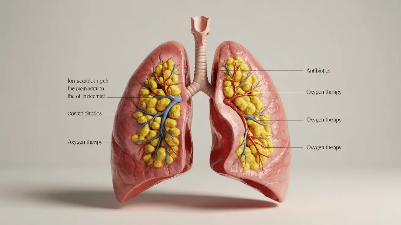
Managing CT Consolidation and Its Impact on Lung Health
Effective management of CT consolidation centers on identifying and treating the underlying cause, while supporting overall lung health and preventing complications. Management strategies may include targeted antimicrobial therapy for pneumonia, diuretic therapy and fluid management for pulmonary edema, or procedures addressing hemoptysis or obstruction when a tumor is involved. Patient education about symptom monitoring, adherence to therapy, and follow-up imaging is essential, as is coordinating care with primary and specialty providers to optimize outcomes in the context of comorbid respiratory diseases.
- Initiate cause-specific treatment promptly (e.g., antibiotics for bacterial pneumonia, supportive care for edema).
- Use follow-up imaging to assess treatment response and detect progression or new complications.
- Address risk factors and comorbid conditions that can worsen lung health, such as heart failure or autoimmune disease.
- Engage in shared decision-making with the patient and caregiver to align care with goals and preferences.
| Diagnostic Step | Rationale |
|---|---|
| Clinical evaluation | Characterizes symptoms and guides differential diagnosis |
| Initial imaging (X-ray) | Detects consolidation and guides need for CT |
| Chest CT | Defines extent, pattern, and possible causes such as infection, edema, hemorrhage, or tumor |
| Laboratory tests | Assists in identifying infectious vs inflammatory etiologies and organ involvement |
| Follow-up imaging | Monitors response to therapy and detects complications |
For readers seeking practical implications for employment or clinical practice considerations in 2025, consult additional resources such as Agency recruitment is transforming the hiring process and How a recruiter helps you find the best jobs to understand how expertise and trust shape outcomes across fields. If you’re curious about technology’s role in operations and industry growth, these articles offer broader context: Robotics transforming logistics operations and Chicago’s industrial growth and key sectors.
In practice, a well-coordinated approach that combines clinical evaluation, imaging, and carefully chosen treatment plans tends to yield the best outcomes for patients with pulmonary consolidation. Clinicians should tailor management to the suspected underlying cause, while maintaining vigilance for potential complications and the impact on long-term lung health.
For more insights into how dedicated professionals support individuals in navigating complex job landscapes while maintaining high standards of care, explore these resources: How a recruiter helps you find the best jobs, Agency recruitment is transforming the hiring process, Robotics transforming logistics operations, and Chicago’s industrial growth and key sectors.
Disclosure: this article was produced with AI assistance and fact-checked against the cited sources.